Enhancing Cancer Therapy Using Functionalized Photosynthetic Bacteria
Researchers succeed in enhancing the anticancer efficacies of purple photosynthetic bacteria by tagging them with bioactive molecules and polyethylene glycol derivatives
Selective targeting of cancerous cells poses major clinical challenges during cancer therapy. However, this limitation can be overcome by using bioengineered bacteria with highly optimized chemical modifications. A recent study demonstrates the use of chemically modified purple photosynthetic bacteria for the successful detection and elimination of colon cancer cells in a mouse model. The study also sheds light on the underlying mechanism of action.
Targeting malignant tumors with high precision is challenging for biomedical researchers. However, this scenario is likely to witness a paradigm shift in the near future, through the use of specially engineered bacteria, that can eliminate malignant cells efficiently.
Using bacteria to target cancer cells, or bacterial therapy, can be further enhanced through genetic engineering and nanotechnology. However, its efficacy may be hindered due to technical constraints and the potential development of antibiotic resistance. Hence, it is crucial to achieve the moderate yet effective chemical modification of bacteria for improved biocompatibility and functionality, such that their medical abilities are not compromised.
Recently, certain types of purple photosynthetic bacteria (PPSB) have come into limelight for their potential to address the challenges of bacterial therapy. Exploring this further, a study published online on 14 August, 2023 in Nano Today reports the use of chemically modified PPSB for detecting and eliminating hard-to-eradicate cancerous cells in a mouse model.
The study, led by Associate Professor Eijiro Miyako from the Japan Advanced Institute of Science and Technology (JAIST), selected Rhodopseudomonas palustris (RP) as the optimal bacterium for conducting the studies. "RP demonstrated excellent properties, such as near-infrared (NIR) fluorescence, photothermal conversion, and low cytotoxicity. It absorbs NIR light and produces free radicals--a property that can be utilized to kill cancer cells," explains Prof. Miyako.
In an attempt to improve the therapeutic efficacy of the isolated strain, the team sought chemical modifications to alter the bacterial membranes. First, they performed membrane PEGylation, or the attachment of polyethylene glycol derivatives to the bacterial cell walls. Prior research indicates that bacterial PEGylation helps in evading host immune response and converts light energy into heat, which can then be utilized to selectively eliminate cancerous cells.
The initial results were encouraging. For instance, coating the RP membrane surface with a "Biocompatible Anchor for Membrane (BAM)" did not adversely affect RP cell viability for at least a week. Moreover, the BAM-functionalized RPs were not eliminated via phagocytosis by macrophages--cells that play a key role in the immune system's defensive actions against bacterial invasions.
Next, the researchers attached a fluorescent "Alexa488-BSA" conjugate to the BAM-functionalized RPs, thus creating a bacterial complex with a trackable fluorescent marker. This conjugate was subsequently replaced with a "PD-L1" antibody. Prior studies have shown that cancer cells express a protein called "Programmed Cell Death Ligand 1 (PD-L1)" on their surface. PD-L1 can smoothly turn off the host defense system by binding to PD-1 receptors. This allows the cancer cells to evade immune detection and elimination. Anti-PD-L1 antibodies block this interaction, thus preventing cancer cells from bypassing immune-system-mediated destruction.
As expected, both anti-PD-L1-BAM-RP and RP, inhibited tumor growth in a murine model of colon cancer. However, anti-PD-L1-BAM-RP, BAM-RP, and RP, when excited with a laser, showed an especially dramatic anticancer effect. In fact, solid tumors vanished completely following the laser irradiation of anti-PD-L1-BAM-RP, BAM-RP, or RP that were injected into tumor-bearing mice. Further, on assessing photothermal conversion properties, both anti-PD-L1-BAM-RP and natural RP exhibited strong photothermal conversion due to the presence of light-driven bacteriochlorophyll (BChl) molecules.
Among the various bioconjugates, anti-PD-L1-BAM-RP showed the highest efficacy in the initial stage of the treatment. Moreover, it was not toxic to surrounding healthy cells or to the murine host. Subsequent experiments revealed the underlying mechanism of colon tumor annihilation in the mouse model.
"Our findings revealed that light-driven functional bacteria demonstrated effective optical and immunological functions in the murine model of colon cancer. Moreover, the NIR fluorescence of the engineered bacterial complexes was used to locate tumors, effectively paving the way for future clinical translation," says Prof Miyako.
He further adds, "We believe that this bacterial technology could be available for clinical trials in 10 years and have positive implications for cancer diagnosis and therapy."
Here's hoping that bacterial therapy helps researchers, oncologists, and patients with cancer with much needed relief.
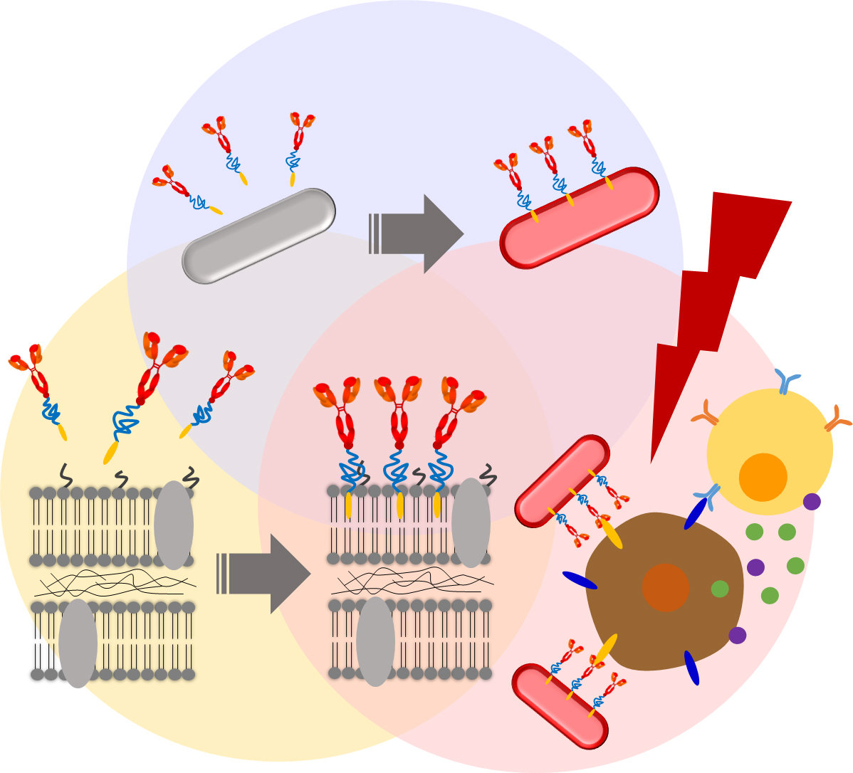
Image Title: Membrane modification of photosynthetic bacteria for enhanced cancer therapy
Image Caption: The membranes of photosynthetic bacteria were PEGylated to improve their biocompatibility and photothermal conversion. Fluorescent markers and an anti-PD-L1 antibody were further attached to enable tumor-targeting and immunological activation. The engineered bacteria demonstrated effective tumor suppression and immunological responses in a mouse model of colon cancer.
Image Credit: Eijiro Miyako from JAIST
Usage Restrictions: Can be reused with attribution
Reference
| Title of original paper: | Cancer immunotheranostics using bioactive nanocoated photosynthetic bacterial complexes |
| Authors: | Sheethal Reghu, Seigo Iwata, Satoru Komatsu, Takafumi Nakajo, Eijiro Miyako* |
| Journal: | Nano Today |
| DOI: | 10.1016/j.nantod.2023.101966 |
Funding information
This work was financially supported by Japan Society for the Promotion of Science (JSPS) KAKENHI Grant-in-Aid for Scientific Research (A) (Grant number 23H00551), JSPS KAKENHI Grant-in-Aid for Challenging Research (Pioneering) (Grant number 22K18440), the Japan Science and Technology Agency for Adaptable and Seamless Technology Transfer Program through Target-driven R&D (Grant Number JPMJTR22U1), Institute for Fermentation, Osaka (IFO), and the Uehara Memorial Foundation.
August 29, 2023
